Nail Fungus Diagnosis
How To Diagnose Dermatophyte Infection?
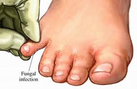
 In order to effectively treat fungal infections of the body especially nail fungus, laboratory tests are essential to confirm diagnosis. But in practice this seldom happens especially if you were to visit a general physician. He would most likely look at the appearance of the nail fungus infection and reach a diagnosis.
Nail fungus infections are mostly treated with medicines that are taken orally. However, for oral medications to be successful, the correct antifungal preparation has to be prescribed. Therefore, the infection has to be confirmed with the help of laboratory tests. In other words, laboratory diagnosis of nail fungus infection could help in the prescription of the right drugs to treat the infection. But the problem is that very few laboratories have the expertise in carrying out these tests.
Why the need for laboratory tests?
The commonest cause of the destruction of the nail plate is nail fungus infection. But nail infection accounts for only half of such cases. Hence there is a need for laboratory verification of diagnosis. For example, if psoriasis is suspected then further tests like nail histology would become necessary. In other words, the cause of destruction of the nail plate should be found out, as for example whether it is because of fungus infection or some other reason.
If the infection is suspected on the basis of its appearance as judged by your doctor but is negative on microscopy and culture then the lab tests should be done once or twice. Then only can fungal infection be ruled out.
Taking the sample for lab analysis
The specimen sample should be taken from the right area of the nail fungus infection.
In the case of a skin infection a skin scrapping from the infected area would be enough as a sample. This sample could be a dry sample or else the scrapings could be taken after soaking the area with saline. The saline method is used to prevent the skin scales from floating in the air and risking infection for others.
In the case of a nail fungus infection what type of sample do you take? Will sample nail scrapings suffice? A nail fungus infection is basically an infection of the nail bed than the nail plate itself. Therefore, the best thing to do is to use a small probe and get a sample of infected skin from under the nail. The most active fungus could be found in the nearest part of the infection. Hence, getting a specimen sample from the nearest part of the infection would be advantageous.
Unfortunately nail clippings are unlikely to have the fungus. That apart, nail specimens are difficult to handle in the laboratory as well. So it is pretty useless sending nail scrapings to the lab other than for superficial white onychomycosis (SWC) infection which is a less common type of nail fungus infection.
What happens when the samples reach the lab?
The samples are treated with potassium hydroxide and examined under a microscope. Thereafter the technician doing the lab tests must have a certain amount of expertise to differentiate fungal elements from the cell walls, dermatophyte from a non-dermatophyte mould and differentiate a dermatophyte from yeast.
If a specimen has to be cultured then it is done in Sabaraud’s dextrose agar medium. If the specimen is alright it will grow in the culture, otherwise not.
Often the specimen sample may have been taken from an area where the infection was old, and most likely the fungus would have been dead. In that case it may not grow in culture. This is the problem with specimen samples taken from nail fungus infections. It could well be that the same specimen would have been positive in microscopy. Therefore direct microscopy is enough for lab confirmation of nail fungus disease.
Microscopic tests would take a few days. Culture results will take 4-6 weeks to materialize.
How are results interpreted?
Dermatophte infection can be confirmed if either microscopy or culture results are positive. But in the case of Candida infections both microscopy and culture have to be positive to confirm disease. In the case of non-dermatophytes both microscopy and culture has to be positive on a minimum of two different samples taken at two different times. In any case, nail fungus infection due to non-dermatophyte moulds are pretty rare.